Elucidate molecular mechanisms of disease
An expansive imaging area to locate rare and meaningful expression events
Cell-type composition and expression of immune response genes are two key variables in neurodegeneration mouse model studies. Single-cell RNA sequencing provides a powerful approach to study these variables, at the expense however of critical anatomical and morphological information. Here we present a methodology which allows accurate gene expression quantification without compromising spatial resolution.
The Rebus Esper™ automated high-throughput spatial omics platform validated the expression of 30 target genes at the single-cell level in the hippocampus of two disease models plus a wild-type control mouse. Using these 30 genes and two proteins of interest, the anatomical structures, major neuronal and glial cell types, and changes in disease-related genes could be visualized and analyzed.
Visualizing changes in the relative abundance of defined cell types and the relationship between these pathological gene-expression levels and the presence of disease-related proteins demonstrate the ability of the Rebus Esper spatial omics platform to yield high-throughput spatial omics data with single cell resolution.
The combination of large imaging area, high-plex RNA quantification, and protein imaging offers a unique and powerful tool to explore complex tissues with an unprecedented level of detail.
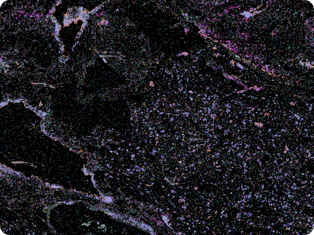
A section of adult human brain processed with the Rebus Esper and Esper High Fidelity Assay reveals the organization of blood vessels in a stroke patient
We were able to define the major neuronal and glial cell types, anatomical structures, and quantify expression changes in disease-related genes.